Brainstorming activities in the classroom
| Õpikeskkond: | Bios4You |
| Kursus: | (7) Neurons: biological and physical model of electrical signal transmission |
| Raamat: | Brainstorming activities in the classroom |
| Printija: | Guest user |
| Kuupäev: | teisipäev, 25. august 2026, 05.55 AM |
Brainstorming activities in the classroom
Guiding phrase: All the activities we perform and all the emotions we feel depend on the transmission of the nerve signal between neurons. How can organic matter collect, analyse and transmit information with very high precision and speed? Our well-being is based on the proper functioning of every element of the neuronal network. What technologies can be used to intervene if something is not going right?
Neurons
What is the structure of the neuron?
The human brain contains about 100 billion cells, neurons, specialized in the transmission of nerve signals, mainly of an electrical nature. A neuron is made up of several parts, with specific functions: a cell body (or soma), containing the nucleus, dendrites and axon. The nerve signal, coming from other neurons or from the receptor organs is collected and transmitted to the nucleus through the dendrites, short and branched extensions of the nucleus. Inside the cell body, the signal is processed and transmitted to the axon, which is a very long extension with the task of carrying the electrical signal to other cells. Separating the inside of the neuron from the external interstitial fluid is the cytoplasmic membrane. Axons are sometimes enveloped for most of their length by a thick insulating material, the myelin sheath. In its final part, the axon branches into a variable number of endings, the synaptic buttons, where the electrical signal is converted into a chemical signal, through a neurotransmitter.
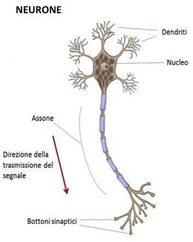
Source image: Online Medicine
What happens when a neuron is not stimulated? What physicochemical characteristics allow us to describe the behaviour of the neuron?
Neurons have a difference in electrical potential between the inside and outside of the cytoplasmic membrane, mainly due to a different concentration of sodium Na+ and potassium K+ ions on the two sides of the membrane. When the neuron is at rest, i.e. it does not transmit signals, this potential difference, called resting potential, has values between -60 mV and -80 mV. The cytoplasmic membrane has ion channels and pumps that open and close in response to certain stimuli, regulating the passage of ions and consequently changing the electrical potential.
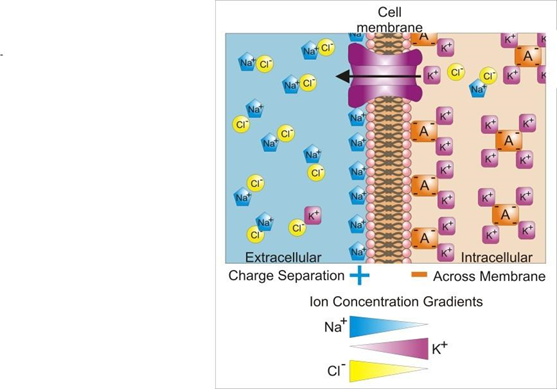
The inside of the cell is rich in K+ ions and negative ions bound to organic macromolecules, while the outside of the cell is rich in Na+ and Cl- ions. The concentration of Na+ ions and K+ ions is regulated by three membrane proteins called ion channels or pumps:
- sodium channels;
- potassium channels;
- the sodium-potassium pump.
The neuron's membrane has many potassium channels: these channels push K+ ions from inside the membrane to the outside, although in this way they increase negativity inside the cell. On the other hand, negative ions inside the cell cannot diffuse outside because they are too large to cross the membrane.
Sodium channels are few and tend to diffuse sodium ions inwards.
The sodium-potassium pump acts against the natural direction of ion flow, pushing Na+ out and K+ inside the cell. When at rest, the membrane is practically impermeable to the ingress of Na+ ions.
 |
Let's think about it together: Neurons are the oldest cells in the body: they are unable to duplicate themselves and in the event of lesions that lead to their death, those around them are unable to replace them. As the years go by, the functionality of the Nervous System becomes less effective: research is therefore interested in finding new methods to enhance the brain's ability to produce new neurons (neurogenesis) to improve learning and memory. What can you do to keep your neurons healthy? How can you keep your mind trained? |
The genesis of the nerve stimulus
How do neurons communicate?
All signals from the outside are picked up by the dendrites and transferred to the nucleus. If a stimulus arrives at the nucleus of a neuron that can modify the resting potential up to a threshold value (about -50 mV), an action potential or nerve impulse is generated that propagates along the axon.
The genesis of the action potential is linked to the exchange of ions through some voltage-gated channels of sodium Na+ and potassium K+. Sodium channels open first, when the potential reaches the threshold value, pushing ions inside the cell that is negative and rapidly depolarizing the membrane. The flow of sodium ions inside the membrane causes a polarity reversal: inside the membrane there is now an excess of positive charge, and outside a lack of positive charge. In this way, the membrane depolarizes, until it assumes a potential of +50mV, the maximum value of the action potential.
The depolarisation condition, however, lasts from 1 to 2 milliseconds.
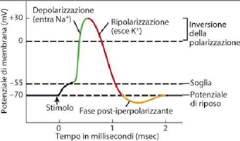
After this Over time, the voltage-gated sodium channels close and the voltage-gated potassium channels open. The latter open more slowly and remain open longer: the K+ ions flow to the outside of the membrane which begins to repolarize, bringing the potential back to a negative value. The flow of K+ persists generating a phase of hyperpolarization and the potential becomes more negative than the resting potential. Finally, all voltage-gated channels close and the activity of the sodium-potassium pump resumes, which restores the resting potential of the plasma membrane. The amplitude (about + 50 mV) and the duration (about 2 milliseconds) of the action potential are fixed, i.e. they do not depend on the amount of excitation.
How does the action potential propagate along the axon?
The electrical signal that propagates through the neuron and is then transmitted to subsequent neurons performs the following sequence of extremely rapid events:
- the electrical stimulus processed by the nucleus causes the opening of voltage-gated sodium channels in a segment near the nucleus; When the threshold value is reached, the first action potential is generated.
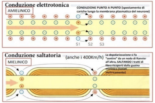
- depolarization propagates along the axon causing the channels to open voltage-gated sodium found in adjacent membrane zones; consequently, a second action potential is generated, which in turn will generate a third and so on.
- At the same time, in the area that had depolarized first, voltage-gated potassium channels opened, bringing the membrane back to the resting potential.
- potentials propagate only in one direction because, after closing, voltage-gated sodium channels undergo a refractory period during which they do not can open again.
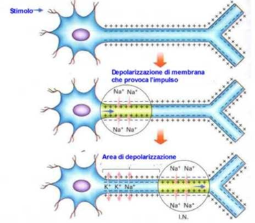
Propagation of action potential along an unmyelinated axon: video at: Propagation of action potential in an unmyelinated axon - Animated medical physiology - YouTube
What role does the myelin sheath play?
Nerve fibres coated with myelin sheath are more waterproof, as the sheath acts as an electrical insulator: consequently, the depolarization and propagation of the action potential occurs only in the sections where it is not present, called Ranvier's nodes. The nerve signal "jumps" from one node to the next with a higher speed than unmyelinated fibres.
 |
Let's think about it together: The transmission of the electrical signal in a neuron first and to other neurons later, is fast and precise. The speed of transmission and the accuracy of information are decisive factors in carrying out all human activities correctly. When something in this wonderful mechanism jams or stops, causing a neurological disease, the impact on the quality of life of affected individuals is devastating. Research on the operating principles of signal transmission neuronal stimulation is the basis of the most advanced neuro-technologies, which exploit electrical stimulation as a therapy. Have you ever heard of electrodes being applied to paralyzed patients who have walked again? |
The physical model of signal propagation in a neuron
What physical model can explain the principle of operation of a neuron?
For a quick refresher on capacitance and capacitors: Capacitors and capacitance of a capacitor (youmath.it)
For a quick refresher on resistance and resistors: Resistors, electrical resistance and electrical resistivity (youmath.it)
The axon is similar to a coaxial cable. The inner part of the axon is the central conducting core of the cable.
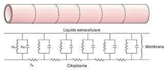
Its strength is high, mainly due to its small cross-section. To describe the electrical properties of the cytoplasm, the resistance per unit length ri is introduced.
The external part of the axon is also a conductor, but the external section is much larger than the internal one and therefore the resistance offered by the interstitial fluid is negligible compared to the internal one. The insulating membrane constitutes a capacitor, since the inside and outside of the axon behave like two conducting plates, storing charges of opposite signs. In parallel with the capacitor, a resistance must be considered, which considers the flow of ions through the membrane itself. To schematize the electrical behaviour of the membrane, two quantities are introduced, the capacitance per unit length cm and the conductivity per unit length of the membrane σμ (φρομ ωηιχη ωε ωιλλ δεριϖε τηε resistance of the membrane per meter rm).
The axon can then be schematized by a circuit consisting of a succession of meshes with resistors and capacitors.
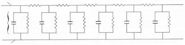
The stimulus potential will depend on the time and location along the axon. In fact, it is characterized by two parameters: the time constant τ and the space constant λ. The mathematical model of the circuit allows us to derive the trend of the potential both as a function of time and as a function of position. In the first case we will have:

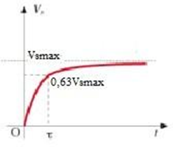
where Vsmax is the maximum value reached by the potential and τ =rmcm.
The time constant τ (on the order of ms) is the time interval it takes for the stimulus potential to reach 63% of its maximum value: it is a measure of how fast the neuron responds.
As we move away from the point where it is generated, the stimulus potential decays with distance due to the combined effects of the ri and rm resistances.
The stimulus potential tends to decrease according to the law:
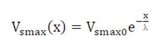
where Vsmax0 is the stimulus potential at the site of origin and λ is the space constant.
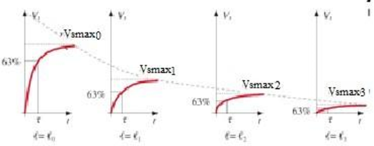
The space constant λ (of the order of mm) is the distance at which the initial potential difference decays to 37% of its maximum value.
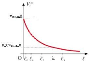
It is a measure of how far the current can flow down the axon before dissipating due to leakage currents.
So after traveling a distance of λ, the signal has reduced to about 1/3 and after 2 λ it is about 1/7 of the original signal and can no longer trigger the action potential. In reality, the stimuli travel considerably longer distances, of the order of a couple of meters, due to the intervention of other mechanisms that we are not considering. Mathematically, the space constant is described as:
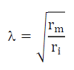
The main factor influencing the internal resistance ri is the radius of the axon: axons of large diameter have a lower resistance to longitudinal current flows and, therefore, a lower axial resistance; consequently, they will have a of the axon ra.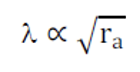
What does the speed of propagation of the nerve impulse depend on?
The knowledge of the two constants allows us to find the speed of propagation of the electrical signal along an axon:
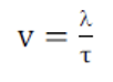 and therefore, to deduce that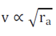
relationship that agrees with the experimental results.
 |
Let's reflect together: The term model is a representation sometimes a simplification of a phenomenon capable of explaining how it works. Using a physical model to interpret and understand a biological phenomenon answers to the need highlighted by several neuroscience scholars of |
Analysis of the model from a mathematical point of view
Flipped Learning
Read the proposed article. Focus especially on the study of nerve signals and electrophysiology techniques described in the article. He then completes the study by analysing which fields of research, according to the article, are most effective for the study of neuronal circuits
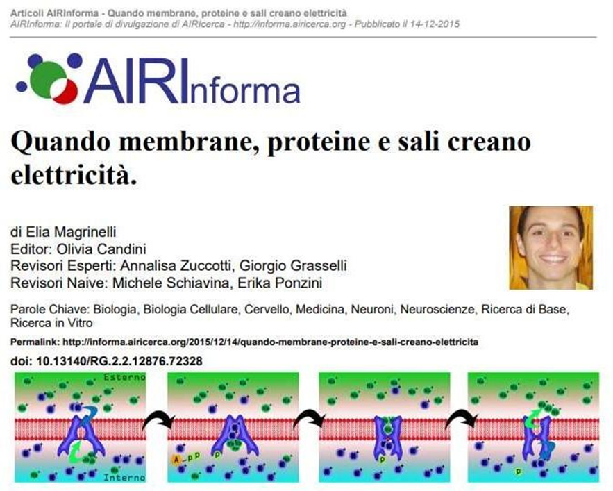
We comment on the study of nerve signals proposed in the article:
Most common electrophysiology techniques:
- The Patch Clamp technique
- The inside-out and outside-out technique
- extracellular recording
Particularly effective alliances with electrophysiology:
- pharmacology
- genetic engineering, optogenetics