Explore Module
| Sito: | Bios4You |
| Corso: | (33) The vision mechanism: from light to the brain |
| Libro: | Explore Module |
| Stampato da: | Svečio paskyra |
| Data: | martedì, 25 agosto 2026, 05:56 |
Engagement phase
Guiding question. Do we see with our eyes or with our brain?
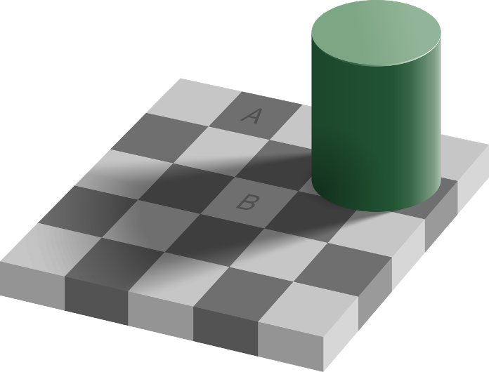
The lesson opens with a brief visual stimulus: a picture in which the same surface appears to have different colors depending on the surrounding context, or a brightness illusion. The purpose is not to “deceive” students, but to introduce the idea that vision is not a mere optical recording—it is an interpretation of physical signals performed by biological structures.

Initial stimuli (videos and short resources)
How the Eyes Work – National Eye Institute (NEI): a clear overview of the main parts of the eye and the light path through the ocular media.
https://www.nei.nih.gov/learn-about-eye-health/healthy-vision/how-eyes-work
Our Eyes – Our Vision – Science Learning Hub: an accessible summary of the neural pathway from retina → optic nerve → brain.
https://www.sciencelearn.org.nz/resources/3006-our-eyes-our-vision
PhET – Geometric Optics (lenses/images): a short interactive exploration about refraction and focusing.
https://phet.colorado.edu/sims/html/geometric-optics/latest/geometric-optics_all.html
Kick-off activity (5–10 min).
A quick projection of an optical illusion or a short video clip invites students to ask, “Why do we see something that isn’t there?”
The goal is to highlight that vision is an interpretative process guided by the brain, not just an optical “input”.
Suggested video: How Vision Works (Academy of Science – Curious)
https://www.science.org.au/curious/video/vision
Brainstorming activities in the classroom
Leading question. Without light, there is no vision: what happens to light as it travels through the eye, and how does it become an electrical signal interpretable by the brain?
Prompting questions
Refraction in the cornea and crystalline lens; the pupil regulates the amount of light; accommodation of the lens maintains sharpness at different distances.
Because the activity of rods predominates (high sensitivity but no color discrimination), while cones require more light to distinguish wavelengths.
Does the retina “record” a ready-made image?
No; the retina performs pre-processing (contrast, movement, direction), then the signal travels to the thalamus and visual cortex, where information is integrated.
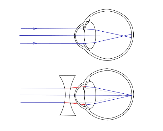
Because the focal plane depends on geometry and refractive indices; when the image plane does not coincide with the retina, the image is out of focus (myopia/hyperopia).
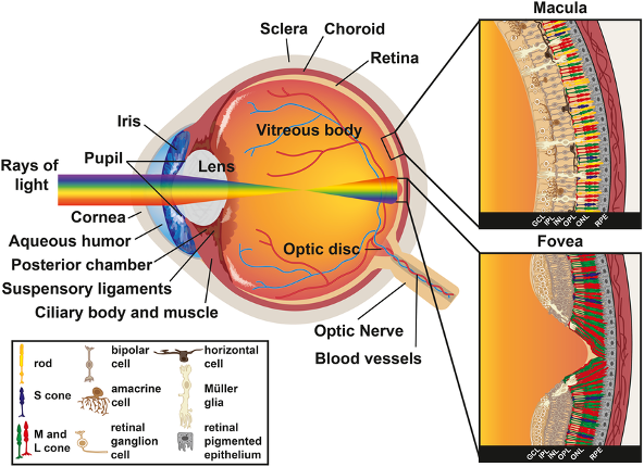
Label the parts of the eye (cornea, pupil, iris, lens, retina, optic nerve) using a worksheet or an interactive resource.
Suggested resource: Labelling the Eye – Science Learning Hub
https://www.sciencelearn.org.nz/resources/3007-labelling-the-eye
Physics and Optics of Vision
Light, refraction, and image formation.
Vision begins when light reflected by objects enters the eye and passes through a natural optical system: cornea, aqueous humor, crystalline lens, and vitreous body. At each interface, light is refracted according to differences in refractive index. The result of this process is the focusing of the image on the retinal plane. The quality of the image depends on three main factors: surface geometry (curvature), refractive indices, and the pupil diameter controlling the depth of field. If light converges before the retina (myopia) or beyond it (hyperopia), the image appears blurred; the crystalline lens compensates through accommodation within physiological limits.
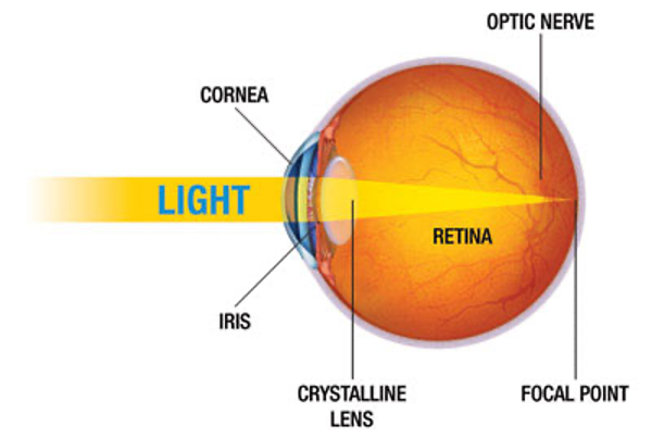
What determines where the focus will fall?
The balance between optical curvature and object distance: the closer the object, the greater the curvature required to maintain a sharp image on the retina.
From physics to physiology: photoreceptors and signal.
On the retina, the inverted image is not a final “snapshot”: it is the physical stimulus that triggers signal transduction. Photoreceptors (rods and cones) contain photopigments that, upon photon absorption, change state and activate a biochemical cascade. The resulting signal is not an “all-or-nothing” action potential, but a graded potential transmitted to bipolar and ganglion cells. The axons of the ganglion cells form the optic nerve, the output channel carrying information to the brain.
Why can we distinguish large shapes in the dark but not colors?
Because rods are more sensitive and active in scotopic (low-light) conditions but cannot detect color; cones, responsible for color and acuity, require higher luminance levels.
Spatial distribution and visual acuity
Cones are densest in the fovea, where visual acuity is maximal and connections are more direct; rods dominate in the periphery, favoring the detection of movement and light variations. This trade-off explains why reading requires direct foveal fixation, while peripheral vision is more effective for motion detection.
Why do letters become less sharp when we fixate on a single one?
Because cone density decreases away from the fovea, and neural convergence increases: sensitivity improves, but spatial resolution decreases.
From the retina to the brain: mandatory stages
The signal leaves the eye through the optic nerve, passes through the optic chiasm (partial decussation), continues in the optic tract to the lateral geniculate nucleus (thalamus), and finally reaches the primary visual cortex (V1). Here—and in higher-order areas—the information is decomposed into features (orientation, spatial frequencies, motion, color) and then recomposed into a coherent perception. The brain does not “see” photons—it reconstructs meaning from neural activity patterns.
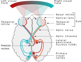
Because perception is generated in the brain: the cortex interprets and reconstructs information regardless of the initial optical orientation.
From theory to everyday objects
The same principles operating in the eye are found in glasses and contact lenses (focus correction), cameras and smartphones (lenses, apertures, sensors), and VR/AR headsets (optics for near-display focusing). Bringing physics out of the lab helps understand not only how a lens works, but when and why optical correction is effective.
Let’s reflect together.
Optimizing the retina is not enough: perception depends on subsequent processing. If the physical light remains the same, why do context and contrast change what we see? What does it really mean to “see with the brain”?
Web-based resources
How the Eyes Work - National Eye Institute (NEI)
https://www.nei.nih.gov/learn-about-eye-health/healthy-vision/how-eyes-work
Interactive explanation of ocular anatomy and light path through transparent media.
Photoreceptors: rods and cones - Khan Academy
https://www.khanacademy.org/science/health-and-medicine/nervous-system-and-sensory-infor/sight-vision/v/photoreceptors-rods-cones
Video lesson on photoreceptors and differences between scotopic and photopic vision.
Geometric Optics - PhET Interactive Simulations, University of Colorado Boulder
https://phet.colorado.edu/sims/html/geometric-optics/latest/geometric-optics_all.html
Interactive simulation: observe how focus and magnification vary with distance and lens curvature; connect results to myopia, hyperopia, and accommodation.
Advanced Level
Fototrasduzione e adattamento.
The cascade occurring within photoreceptors (activation of photopigment, transducin, modulation of cGMP and ion channels) explains how light becomes current. Adaptation (to light and darkness) recalibrates the system’s sensitivity, enabling operation across multiple orders of magnitude in luminance.
Resolution and contrast.
Acuity does not depend solely on optics: retinal and cortical circuits select spatial frequencies and orientations, enhancing contour contrast. The different pathways (parvocellular/magnocellular) prioritize detail/color or motion.
Visual pathways and integration.
Beyond V1, processing splits into networks that emphasize “what” (form, color) and “where/how” (space, action). Color perception emerges from the integration of L/M/S cone responses and chromatic opponencies, while motion perception requires temporal integration and binocular disparity for depth.
Why do we perceive motion better than fine details under low light?
Because in scotopic vision, pathways sensitive to global and temporal variations dominate, with reduced spatial resolution.
Why can a small pupil sometimes improve sharpness?
Because it reduces aberrations and increases depth of field (within the limits of diffraction).
Flipped Learning
Objective. Prepare for classroom discussion by understanding the sequence light → retina → optic nerve → cortex and the physics/biology connections.
Tasks (to be submitted as a 1–2-page report or 6–8 slides):
- Short reading on the anatomy of the eye and the path of light. Highlight: cornea, pupil/iris, lens, retina, optic nerve.
- Worksheet “Cones vs. Rods”: complete a table with sensitivity, role in color/acuity, light conditions (scotopic/photopic), retinal distribution.
- Geometric Optics simulation (thin lens): create two screenshots —
- Focused image (focus on “retina”) with annotated parameters;
- Out-of-focus image (simulated myopia/hyperopia) with notes on how accommodation or corrective lenses restore focus.
- Short synthesis (max 120 words): explain in your own words how the retina does not “record” ready-made images but transduces and pre-processes signals to be sent to the brain.
- Three debriefing questions: one on optics (refraction/focusing), one on biology (phototransduction), one on perception (why don’t we see the world upside down?).
Submission. Upload the PDF or slides to the class channel before the lesson.