1. Explore - Foundations of Viruses, Their Parts and Types
| Website: | Bios4You |
| Kurs: | (13) Understanding Viruses Through 3D Modeling & AR Visualization |
| Buch: | 1. Explore - Foundations of Viruses, Their Parts and Types |
| Gedruckt von: | Επισκέπτης (Guest user) |
| Datum: | Dienstag, 25. August 2026, 06:42 |
Beschreibung
In this section, students build the scientific foundation needed to understand what viruses are and why they are unique biological entities. They explore how viral structure relates to function, how viruses replicate using host cells, and how viruses can be classified into different types. This Explore phase prepares learners for the next stages of the unit, where they will model viruses using 3D tools and visualize them in Augmented Reality (AR), transforming invisible biology into interactive learning objects.
1.1 What is a Virus?
A virus is a microscopic infectious agent that cannot reproduce independently. Unlike living cells, viruses do not have the full machinery needed to produce energy, grow, or divide. Instead, they must enter a host cell and “use” the host’s internal systems to create new virus particles.
Viruses can infect many types of organisms, including animals, plants, fungi, and bacteria. Although simple in structure, viruses can spread efficiently and evolve quickly, which is why they are important in biology, biotechnology, and public health.
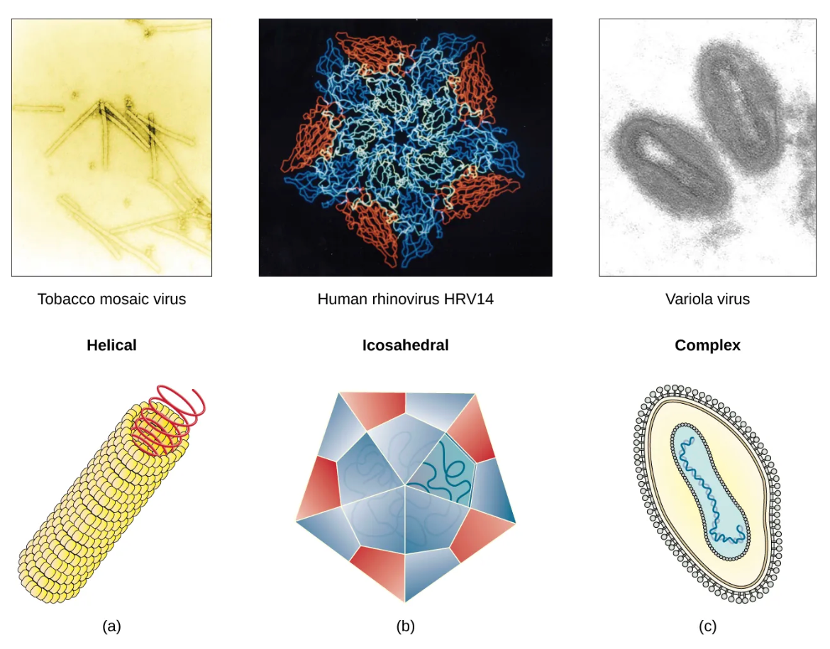
Figure 1. Viruses example. Source:
https://openstax.org/books/biology-2e/pages/21-1-viral-evolution-morphology-and-classification
Example
The influenza virus cannot make proteins on its own. Only after it enters a human cell can it use the cell’s machinery to produce new virus particles. This dependency explains why viruses are inactive outside a host.
1.2 Virus vs. Cell: Key Differences
To understand viruses, it is helpful to compare them with cells:
- Cells are living systems. They have a cell membrane, cytoplasm, and internal structures (organelles in eukaryotic cells). Cells can produce energy and reproduce on their own.
- Viruses are not complete living systems. They do not have cytoplasm, organelles, or metabolism. Outside a host cell, a virus is inactive.
This difference explains why viruses are often described as being on the border between living and non-living: they show life-like behavior only inside a host cell.
Example
A bacterium can live independently in many environments, while a virus infecting that bacterium (a bacteriophage) remains inactive until it finds the correct host.
1.3 The Main Parts of a Virus (Structure)
Even though viruses vary widely, many share core structural components:
- Genetic material (genome): The “instruction code” of the virus. It can be DNA or RNA. Example Influenza – ssRNA (+): https://microbenotes.com/classification-of-virus/
- Capsid: A protective protein shell that surrounds and protects the genome. Capsids often have geometric shapes that help stability and packaging. Example: Adenovirus – icosahedral capsid: https://bio.libretexts.org/Bookshelves/Introductory_and_General_Biology/General_Biology_2e_(OpenStax)/05:_Unit_V-_Biological_Diversity/5.01:_Viruses/5.1.02:_Viral_Evolution_Morphology_and_Classification
- Envelope (in some viruses): A lipid outer layer taken from the host cell membrane. Example: HIV – enveloped with gp120 spikes https://en.wikipedia.org/wiki/Viral_envelope
- Spikes / surface proteins (in many viruses): These help the virus recognize and attach to the host cell. Example: SARS-CoV-2 spike protein: https://morgridge.org/community/teaching-learning/virology-immunology/factsheets/virus-structure/
Key idea for students: Virus structure is not random — each part supports a function (protection, attachment, entry, replication).
To support visual and conceptual understanding of virus structure, students are encouraged to watch a short educational video explaining the basic biology of viruses. The video clearly presents the main parts of a virus, including genetic material, capsid, envelope, and surface proteins, and explains how these components work together during infection. This resource helps learners connect written descriptions with dynamic visual explanations, making complex microscopic structures easier to understand before moving on to 3D modeling and AR activities.
Recommended video resource: Khan Academy – Viruses
https://www.khanacademy.org/science/biology/biology-of-viruses/virus-biology/v/viruses
1.4 How Viruses Infect Cells (Viral Life Cycle – Basics)
A simplified viral life cycle includes these stages:
- Attachment: The virus binds to specific receptors on the host cell surface using spikes or surface proteins.
- Entry: The virus (or its genome) enters the host cell. Enveloped viruses often fuse with membranes; others may enter through endocytosis.
- Replication & protein production: The host cell produces viral genetic copies and viral proteins.
- Assembly: New virus particles are assembled from newly made parts.
- Release: New viruses leave the cell, either by bursting the cell (lysis) or by budding (common in enveloped viruses).
This life cycle explains why viruses depend on hosts and why they can cause damage: viral replication can disrupt normal cell function.
1.5 Virus Types: How Can We Classify Viruses?
Viruses can be grouped in different ways. For this unit, students focus on classifications that connect clearly to modeling and AR visualization:
- By genome type: DNA viruses (herpesvirus, etc.) vs. RNA viruses (influenza, etc.). (More: https://pmc.ncbi.nlm.nih.gov/articles/PMC7150055/)
- By envelope: Enveloped (HIV, influenza, etc.) vs. non-enveloped (poliovirus, norovirus, etc.). More: https://virologyresearchservices.com/2022/05/22/enveloped-vs-non-enveloped-viruses/
- By shape: Helical (tobacco mosaic virus – for plants, etc.), icosahedral (adenovirus, etc.), complex (bacteriophage, etc.). More: https://bio.libretexts.org/Bookshelves/Introductory_and_General_Biology/General_Biology_2e_(OpenStax)/05:_Unit_V-_Biological_Diversity/5.01:_Viruses/5.1.02:_Viral_Evolution_Morphology_and_Classification; https://microbenotes.com/classification-of-virus/)
- By host: Animal viruses (HIV, etc.), plant viruses (TMV, etc.), bacteriophages (T4 phage, etc.). More: https://microbenotes.com/classification-of-virus/
These categories are useful because they connect directly to what students will create in 3D: different shapes, different outer layers, different “surface features.”
1.6 Why Do Viruses Matter in Biotechnology and Society?
Viruses are often associated with disease, but their importance extends far beyond causing infections. In modern science and biotechnology, viruses are also used as tools to study biology, develop medical solutions, and understand natural systems.
One important application of viruses is in vaccines and immune science. Scientists study viral surface proteins to understand how the immune system recognizes a virus. For example, research on influenza viruses has shown that small changes in viral surface proteins can affect how well vaccines work each year. Similarly, understanding the structure of viral proteins has helped scientists design vaccines that train the immune system to respond more effectively to infection. These examples show why knowing virus structure is essential for protecting human health.
Connection to 3D modeling: When students design spikes or surface proteins on their virus models, they are representing the same structures scientists study when developing vaccines. The size, position, and number of spikes in a 3D model help explain how a virus recognizes and attaches to host cells.
Viruses are also used in gene therapy and biological research. In laboratories, modified viruses are commonly used as vectors to deliver genetic material into cells. For instance, harmless versions of viruses are used to introduce new genes into cells so scientists can study how those genes function. In some medical applications, viral vectors are used to deliver therapeutic genes to specific cells, demonstrating how viruses can be redesigned for beneficial purposes.
Connection to 3D modeling: By creating a capsid around the viral genome in a 3D model, students visualize how genetic material is packaged and protected. Modeling the capsid as a geometric structure helps learners understand why shape and symmetry are important for stability and function.
Beyond medicine, viruses play a key role in evolution and ecosystems. In marine ecosystems, viruses infect microorganisms such as bacteria and algae, controlling their population sizes. This process helps regulate nutrient cycles in oceans, which are important for global climate balance. Viruses can also transfer genetic material between organisms, contributing to genetic diversity and long-term evolution.
Connection to 3D modeling: When students compare different virus types—such as enveloped and non-enveloped viruses—they can model how structural differences affect survival in different environments. For example, adding or removing an envelope in a 3D model helps explain why some viruses are more resistant to environmental conditions than others.
In this learning unit, viruses are used as a clear example of how real scientific knowledge can be transformed into simplified digital representations. By designing virus structures using basic geometric shapes and visualizing them through Augmented Reality, students experience how scientists and engineers use models to study complex systems. This approach connects biology with design, technology, and STEM skills, preparing learners to understand how biological structures inspire innovation in the real world.
Questions for Reflection
- Why are viruses considered different from living cells?
- How does a capsid protect the viral genome, and why might its shape matter?
- What is the difference between enveloped and non-enveloped viruses, and how might that affect transmission or stability?
- How does “structure → function” help explain why viruses infect specific host cells?
- Which virus feature do you think would be easiest to represent in a 3D model, and which would be hardest? Why?