
Introduction. The Vision Mechanism: From Light to the Brain
Biology of vision and optics; signal transduction; from the eyeball to the visual cortex
Vision arises from the intersection of physical phenomena and biological processes. Light reflected from objects passes through the transparent media of the eye, is refracted, and focused on the retina. Here, photoreceptors convert luminous energy into electrical signals that travel along the optic nerve to the cortical areas responsible for visual processing. Seeing does not mean recording an image passively, but reconstructing it from signals that the brain interprets and integrates.
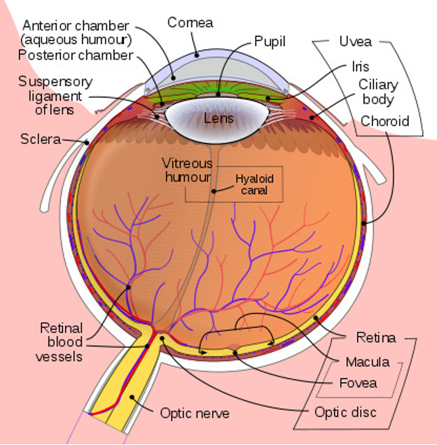
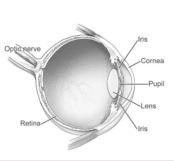
Focusing depends on the curvature of the surfaces and on the refractive indices of the ocular media. Geometrical or optical alterations lead to conditions such as myopia or hyperopia, in which the focal point falls in front of or behind the retinal plane. The crystalline lens contributes to accommodation, slightly changing its curvature to maintain sharpness at different distances.
The retina is a specialized neural tissue. Rods, which are highly sensitive, support vision under low light conditions; cones, less sensitive, enable color perception and sharp vision under daylight. Phototransduction begins when photopigments absorb photons and trigger a biochemical cascade that modulates the membrane potential of photoreceptors and subsequent retinal neurons (bipolar, ganglion, and interneurons).
From the retina, the axons of the ganglion cells form the optic nerve. The fibers partially cross at the optic chiasm, ensuring that the right visual field is processed mainly by the left hemisphere and vice versa. The signal proceeds along the optic tract to the lateral geniculate body (thalamus) and then reaches the primary visual cortex (V1), where, together with associative areas, it is analyzed and integrated to extract features such as shape, depth, motion, and color.
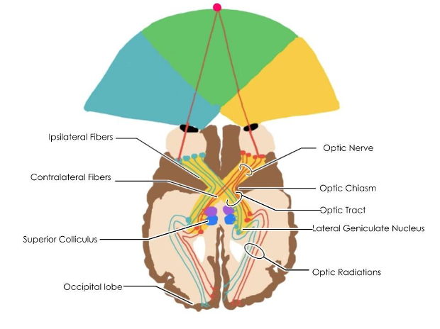
Perception is influenced by mechanisms of adaptation and by inferential processes. Adaptation allows the visual system to maintain sensitivity across a wide range of luminance levels, while visual illusions show that the brain interprets stimuli based on context and experience, completing partial information to reconstruct a coherent scene.
Binocular vision provides disparity cues essential for depth estimation, while monocular cues—such as relative size, perspective, occlusion, and shading—contribute to the three-dimensional perception even when only one eye is used.
From an educational point of view, it is crucial to clarify the sequence of events: (1) light is focused on the retinal plane; (2) photoreceptors convert light into electrical signals; (3) the retinal network pre-processes the signal; (4) the signal travels along the optic nerve and thalamic stations; (5) the visual cortex integrates the information to produce a perceptual representation. Presenting these steps in an ordered way helps students connect physical causes and biological effects.
The targeted use of digital and augmented reality tools makes it possible to visualize otherwise invisible processes, such as the path of light through ocular media, the activation of photoreceptors, and the transmission of signals along the visual pathways. Scene-by-scene visualization facilitates the understanding of relationships between anatomical structures and physical principles.
Why do we not see in the absence of light? How do the curvature of the crystalline lens and the refractive indices of ocular media affect image sharpness? How much does cortical interpretation contribute to our perception of color and motion? These questions help link observable phenomena to the underlying mechanisms.