Exploring Brain Activity Through Augmented Reality
Technology is now ingrained in education, and the results show that it positively impacts learning and teaching methods (Dübel et al. 2014). AR/VR technology can provide simulated experiences that temporarily or permanently replace some components of hands-on instruction (Bakharia et al. 2016). AR provides an efficient way to represent a model that needs visualization so that a brain model can benefit the most from AR. 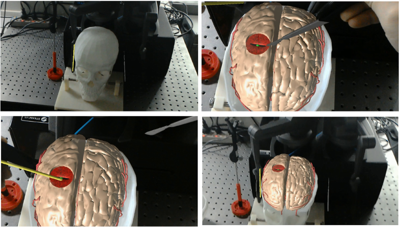
Figure 10. Augmented reality guidance for neurosurgery, source: Si et al., 2019.
In recent years, augmented reality (AR) has emerged as a transformative tool in education and neuroscience, offering new possibilities for visualizing and interacting with complex biological systems. One promising application lies in exploring brain activity, where AR can bridge the gap between abstract neural processes and tangible learning experiences. AR enables real-time, spatial visualization of anatomical structures and brain functions, enhancing learners’ engagement and comprehension (Bacca et al., 2014; Radu, 2014). In the context of neuroscience education, especially among younger learners, AR applications such as Brainapse present an innovative approach to understanding neural mechanisms by overlaying digital brain models with animations of brain activity and cognitive functions.
This aligns with findings by Kucuk et al. (2016), who reported that AR-based learning environments significantly improve students’ conceptual understanding and motivation in science subjects. Furthermore, research by Billinghurst et al. (2015) highlights AR’s potential to support embodied learning, where users actively engage with spatial and temporal representations of brain functions. By integrating AR into neuroscience education, we aim to investigate how these technologies can enhance students’ ability to grasp fundamental concepts in brain anatomy and cognitive processes and foster more profound interest in STEM-related fields.
Brainapse is an interactive educational app that uses augmented reality (AR) to bring the human brain to life. By merging 3D anatomical models with real-world environments, the app offers students an engaging way to explore brain structures, neural pathways, and cognitive functions. It transforms abstract biological concepts into tangible learning experiences, making it especially valuable for younger learners or those new to neuroscience. With its visual clarity and immersive design, Brainapse opens new opportunities for interactive, curiosity-driven learning both in and outside the classroom.
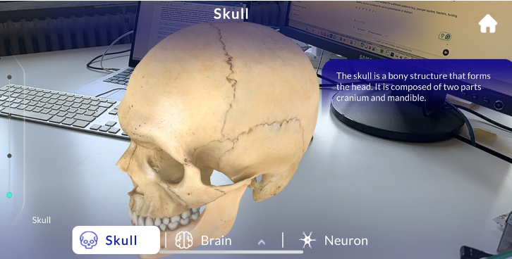
The image below demonstrates the use of augmented reality to visualize the human skull in three dimensions. Through Brainapse, students can explore the anatomical structure of the skull, which consists of the cranium and the mandible. This interactive visualization helps learners understand the skull's protective role and its importance in housing the brain, enhancing spatial awareness and memory retention through immersive technology.
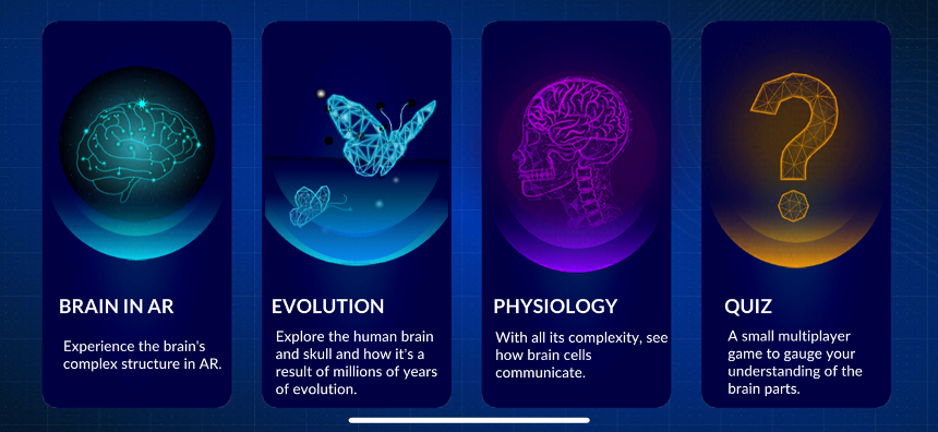
Figure 11. Brainapse App
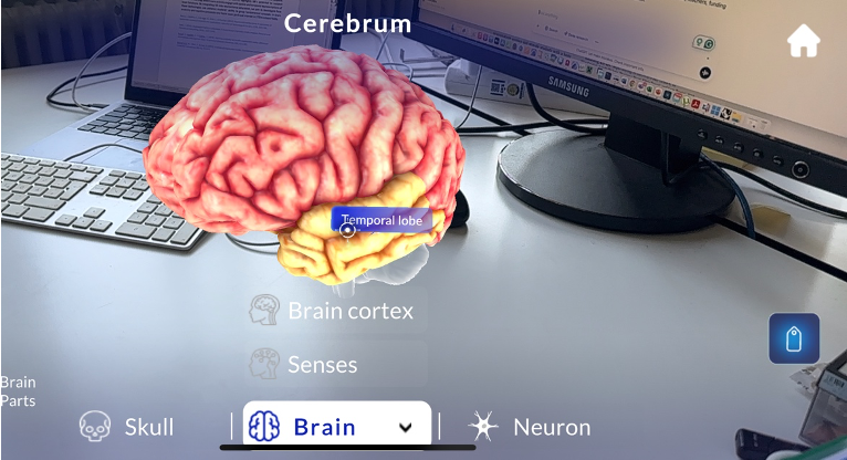
This view below displays the cerebrum focusing on the temporal lobe, a region essential for processing auditory information and memory formation. Using AR, students can directly interact with the labeled parts of the brain, fostering a deeper understanding of functional neuroanatomy. Such interactive elements have improved knowledge retention and learner motivation in science education (Kucuk et al., 2016).
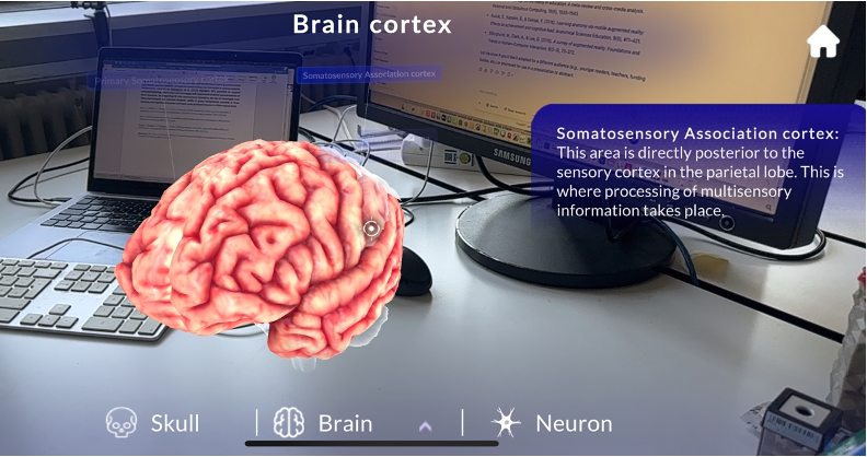
The app highlights the somatosensory association cortex located in the parietal lobe. This region integrates and interprets sensory information from various modalities. Augmented reality makes this concept more concrete for students by presenting it in a real-world context, allowing for experiential learning that supports multisensory engagement (Bacca et al., 2014).
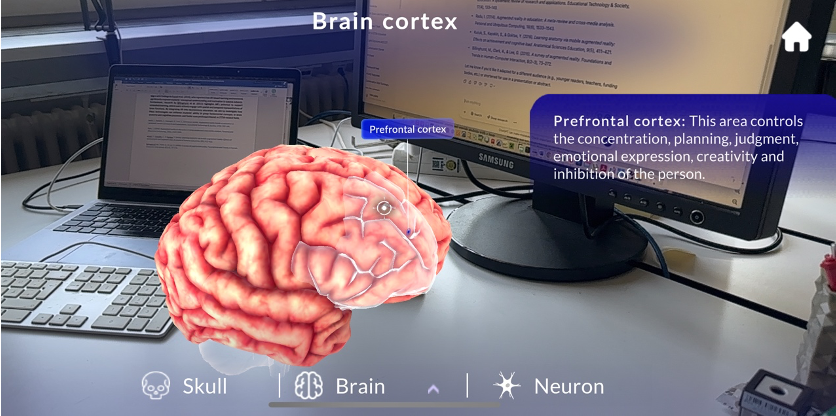 This visualization zooms in on the prefrontal cortex, known for its role in planning, decision-making, creativity, and emotional regulation. Brainapse enables learners to connect cognitive functions with their anatomical locations, creating a powerful learning experience that links biology with behavior, supported by studies in AR-enhanced science learning (Billinghurst et al., 2015).
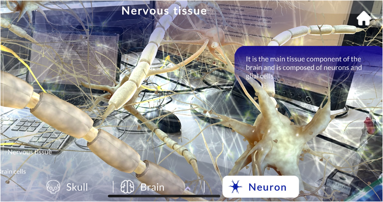
This scene immerses students in a microscopic view of nervous tissue, illustrating neurons and glial cells in a dynamic, 3D environment. By virtually stepping into the brain's cellular landscape, learners appreciate the complexity of neural communication and tissue organization, an experience difficult to replicate through textbooks alone (Radu, 2014).
The Brainapse app offers a unique opportunity to support the learning objectives of this training unit, Light and the Brain: Exploring Neurons and Light Sensitivity, by visually guiding students through the brain’s structure, functions, and neural components. Although the app doesn’t simulate light sensitivity directly, it provides a clear and interactive visualization of key brain areas involved in sensory processing, such as the occipital lobe (responsible for visual perception) and neuronal networks (responsible for transmitting signals, including those related to light stimuli).
By exploring these parts in 3D, students can better understand how neurons communicate sensory information, how light stimuli are processed, and how different brain regions are activated in response. This spatial and immersive learning aligns with evidence suggesting AR enhances conceptual understanding and memory (Bacca et al., 2014; Radu, 2014). Additionally, seeing neurons and nervous tissue in detail fosters a deeper appreciation for how the brain detects, processes, and responds to external stimuli like light, supporting the development of both scientific knowledge and curiosity.